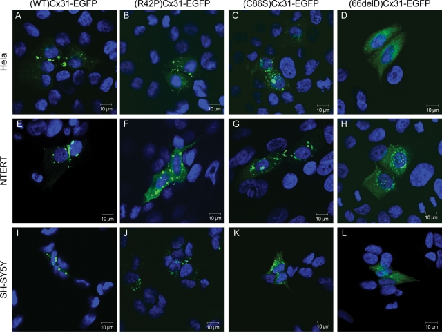
Figure 1.
Distinct subcellular localization patterns are observed upon expression of WT, skin disease- and neuropathy-causing Cx31 in HeLa, NTERT and SH-SY5Y cells. In HeLa cells (A–D), bright aggregates are observed at cell–cell boundaries in cells expressing (WT)Cx31-EGFP (A) which are indicative of gap junction plaques. These aggregates are not observed upon overexpression of any mutant, consistent with their inability to traffic properly to the plasma membrane. Cells expressing either of the skin disease mutants (R42P)Cx31-EGFP (B) or (C86S)Cx31-EGFP (C) display larger brighter aggregates than cells expressing the neuropathy mutant (66delD)Cx31-EGFP (D), which show protein residing on smaller diffuse punctate structures (distinct from the diffuse non-punctate EGFP localization (see Supplementary Material, Fig. S1). A similar subcellular localization is observed when the WT and mutant Cx31 constructs are expressed in the immortalized keratinocyte cell line NTERT (E–H) and the neuroblastoma cell line SH-SY5Y (I–L). DAPI-stained nuclei are shown in blue.

