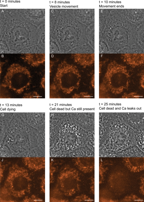
Figure 4.
Calcium Orange does not leave the cells expressing EKV-associated Cx31, while they are undergoing cell death, eliminating a hemichannel mechanism. Examples of images from time-lapse recording of keratinocytes expressing (G12D)Cx31-EGFP. Although EGFP was also recorded, only phase contrast and Calcium Orange images are presented. Time after microinjection is indicated. Cells were loaded with Calcium Orange 30 min after microinjection and recording started 50 min after microinjection. The cell in the centre of the field died 81 min after microinjection and lost calcium 85 min after microinjection. Scale bars are 10 µm.

