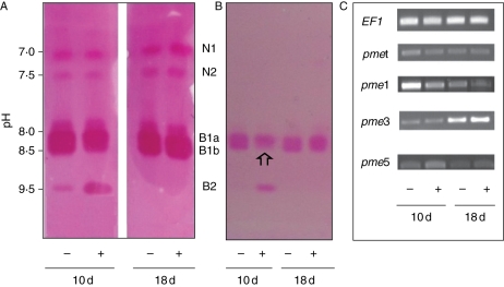
Fig. 2.
Pectin methylesterase isoenzymes ionically bound to the cell wall of the hypocotyl. Seedlings were grown in the presence (+) or absence (−) of cadmium and harvested at 10 and 18 d old. (A) IEF and (B) transfer of the gel onto a pectin–agarose gel. PME activity was revealed by ruthenium-red staining de-esterified homogalacturonans. A similar amount of PME (0·5 nkatal) activity was loaded per well. The pH values measured with a surface electrode are given on the left. Note the decrease of B1b (arrow) which paralleled the increased of B2 at day 10. (C) Expression of Lupme genes compared with the elongation factor gene LuEF1 α (EF1) as seen by PCR. All gel figures were representative of three to five repeats. N and B are neutral and basic isoforms, respectively.

