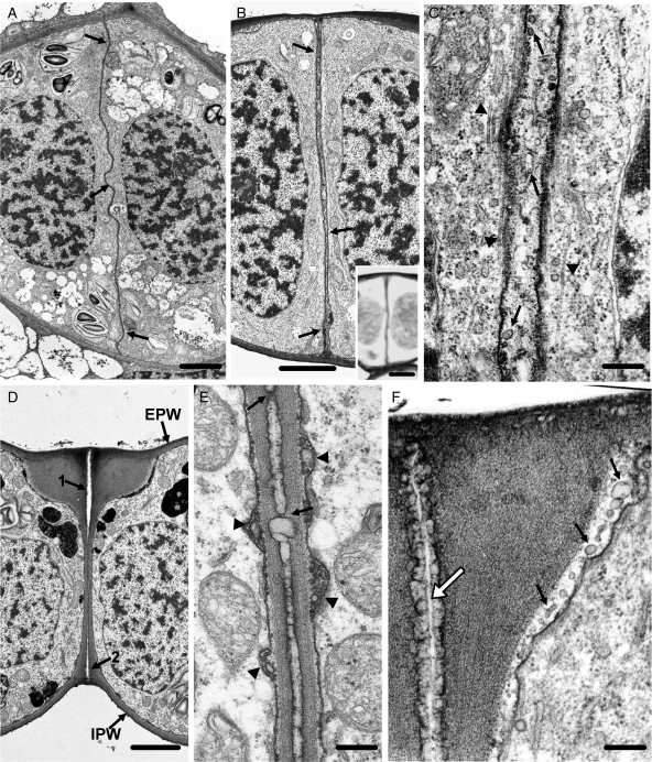
Fig. 12.
TEM micrographs of CPA-affected stomata, in which internal stomatal pore formation has been inhibited. Treatments: (A–F) 25 µm CPA for 24 h. (A) Paradermal view through the middle of a post-cytokinetic affected stoma. The ventral wall (arrows) is wavy and lacks an internal stomatal pore (cf. Fig. 5A). Scale bar = 20 µm. (B) Median transverse section of an affected stoma at a stage of differentiation similar to that of the stoma depicted in Fig. 5B. The ventral wall (arrows) is atypically thickened and lacks an internal stomatal pore (cf. Fig. 5B). Inset: transverse semi-thin section of an affected stoma at a stage of differentiation similar to that of the stoma shown in (B), after PAS staining. The ventral wall and the periclinal walls are positively stained. Scale bars: (B) = 20 µm; inset = 5 µm. (C) Higher magnification of the median region of the ventral wall of the stoma shown in (B). The arrows show membranous elements at inner positions of the aberrant ventral wall and the arrowheads the microtubules. Scale bar = 250 nm. (D) Median transverse section of an affected stoma that is at a stage of differentiation more advanced than that of the stoma shown in (B). The arrows mark the initiating fore- (arrow 1) and rear- (arrow 2) chambers of the stomatal pore. EPW, external periclinal wall; IPW, internal periclinal wall. Treatment: 25 µm CPA for 24 h. Scale bar = 20 µm. (E) The median region of the ventral wall of the stoma shown in (D) at higher magnification. Note the absence of the internal stomatal pore (cf. Fig. 5C, D) and the material localized at the middle lamella. Wall bridges connect the adjacent ventral walls (arrows) and membranous elements are localized in the apoplast (arrowheads). Scale bar = 250 nm. (F) Wall thickening at the junction of the ventral wall with the external periclinal wall of a young affected stoma. The large arrow marks the initiated fore-pore chamber that is lined by a material similar to that localized in the middle lamella in (E). Note the membranous elements in the apoplast (small arrows). Scale bar = 250 nm.

