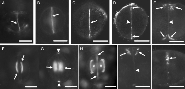
Fig. 3.
Callose localization in developing control stomata after immunolabelling (A, C–J) or aniline blue staining (B). Scale bars = 10 µm. (A) Cytokinetic guard cell mother cell. The arrow indicates the cell plate. (B) Post-cytokinetic stoma. The nascent ventral wall (arrow) exhibits intense callose fluorescence. (C, D) Single CLSM sections through the junction of the ventral wall with the external periclinal walls (C) and a median plane (D) of a stoma in which the internal stomatal pore has been initiated. The margins of the ventral wall (arrows in C and D) exhibit intense callose fluorescence and a less intense one at its median region (arrowhead in D). (E) Median transverse semi-thin section of a stoma, in which stomatal pore formation and wall thickening deposition have started. Wall thickenings containing callose emerge at the junctions of the ventral wall with the periclinal walls (arrows; cf. Fig. 5B). The rest of the ventral wall (arrowhead) lacks callose. (F, G) Optical sections through a surface (F) and an inner plane (G) of a stoma at an early stage of wall thickening. Callose impregnates the wall thickenings at the stomatal pore region (arrows in F and G) and at the junctions of the ventral wall with the dorsal walls (arrowheads in G). (H) Stoma at a more advanced stage of differentiation than that shown in F and G. Callose is detected at the margins of the wall thickenings (asterisks) at the stomatal pore region. The arrows point to ventral wall regions displaying autofluorescence (cf. Fig. 2C). (I) Median transverse semi-thin section of a stoma at the same differentiation stage as that of the stoma shown in (H). Callose is located at the margins of the wall thickenings (arrows). The rest of the ventral wall lacks callose (arrowhead). (J) Transverse semi-thin section of a ventral wall polar end (see Fig. 1A). Callose (arrow) is deposited at the wall thickening developing in this area (cf. Fig. 5E).

