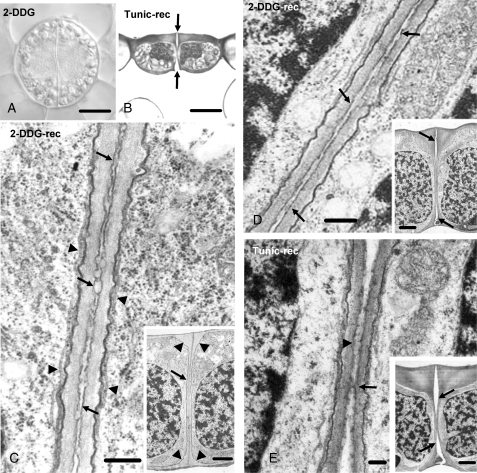
Fig. 8.
(A) Differential interference contrast image of a 2-DDG-affected differentiating stoma. Treatment: 1 mm 2-DDG for 5 d. Scale bar = 10 µm. (B) Median transverse semi-thin section of a tunicamycin-affected stoma recovering under control conditions. This stoma, as well as those illustrated in (C–E), after treatment was placed in distilled water. The arrows point to the forming stomatal pore. Treatment: 12 µm tunicamycin for 5 d; recovery 7 d. Scale bar = 10 µm. (C) Higher magnification of a median transverse ventral wall section of a 2-DDG-affected stoma (inset), recovering under control conditions. The arrows in (C) mark the differentiated middle lamella, while the arrowheads mark microtubules. The arrow in the inset indicates the ventral wall and the arrowheads the wall thickenings. Treatment: 500 µm 2-DDG for 5 d; recovery 7 d. Scale bars: (C) = 250 nm; inset = 2·5 µm. (D) Median transverse ventral wall section of a 2-DDG-affected stoma (inset) recovering under control conditions. This stoma is at a more advanced stage of differentiation than that of the stoma shown in (C, inset). The fore- and rear-chamber of the stomatal pore have been formed (arrows in inset). The arrows in (D) mark the differentiated middle lamella. Treatment: 500 µm 2-DDG for 5 d; recovery 7 d. Scale bars: (D) = 250 nm; inset = 2·5 µm. (E) Median transverse ventral wall section of a tunicamycin-affected stoma (inset) recovering under control conditions, which is at a more advanced stage of differentiation than that of the stoma shown in (D, inset). The stomatal pore is at final stages of formation. The fore- and rear stomatal pore chambers (arrows in inset) have expanded towards the middle of the ventral wall. Note the material localized at the region of ventral walls that have not yet been separated (arrowhead in E). The arrow in (E) indicates a wall bridge connecting the adjacent ventral walls. Treatment: 12 µm tunicamycin for 5 d; recovery 7 d. Scale bars: (E) = 125 nm; inset = 2·5 µm.

