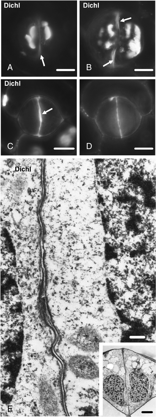
Fig. 9.
Dichlobenil-affected stomata as they appear after aniline blue staining (A–D) or under TEM (E). Treatments: (A–E) 100 µm dichlobenil for 48 h. (A, B) Affected stomata at successive stages of differentiation. Atypical callose depositions can be seen at various sites of the periclinal walls. The arrows show regions of the ventral wall exhibiting autofluorescence (cf. Fig. 2B). Scale bars = 10 µm. (C, D) Optical sections through an external (C) and a median plane (D) of an affected stoma, which is at a stage of differentiation similar to that of the stoma shown in Fig. 3C and D. The external (C) and the median (D) region of the ventral wall emit intense callose fluorescence. The arrow in (C) indicates the initiating wall thickening at the junction of the ventral wall with the external periclinal wall. Scale bar = 10 µm. (E) Higher magnification of the median ventral wall region of the stoma shown in the inset. The internal stomatal pore has not been formed (cf. Fig. 5B). Inset: median transverse section of an affected stoma, which is at a stage of differentiation similar to that of the stomata shown in Fig. 5A and B. Note the absence of the wall thickenings at the sites of junction of the ventral wall with the periclinal walls (cf. Fig. 5B). Scale bars: (E) = 250 nm; inset = 5 µm.

