
FIGURE 4.
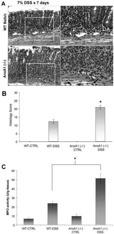
AnxA1-deficient mice have more severe histopathologic injury in DSS-induced colitis. Representative photomicrographs of H&E-stained histologic sections (A) demonstrate more severe colonic injury in AnxA1 (−/−) animals compared with WT controls following 7 days of DSS treatment. Note the increased degree of epithelial injury, increased leukocyte infiltration, and involvement of submucosal tissues in AnxA1 (−/−) mice (inset, arrows). Histologic scoring of colonic injury (B) is significantly higher than that of WT DSS-treated animals (*, p-value <0.05, n = 7 mice). Colonic MPO activity (C) is also significantly higher in AnxA1 (−/−) mice compared with WT BALB/c animals following DSS treatment (*, p-value <0.05, n = 7 mice).