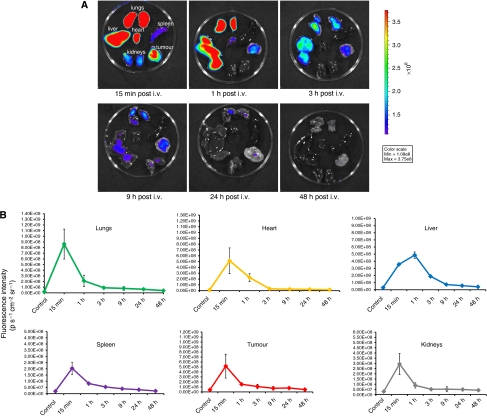
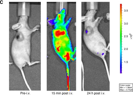
Figure 3.
In vivo biodistribution of ADPM06 as measured by optical imaging. (A) Representative images of excised organs from Balb/C nude mice bearing subcutaneous MDA-MB-231-luc tumours. Mice were killed over a 48-h interval after i.v. injection of ADPM06 (2 mg kg−1). Fluorescence intensity peaked in all organs 15 min post injection, with the exception of the liver, which reached maximum fluorescence at 1 h. Fluorescence intensity reached baseline levels by 24 h and seems to be cleared from the animal by 48 h. MDA-MB-231-luc tumour model used in biodistribution studies to prevent GFP autofluorescence into the NIR channel. (B) Quantification of fluorescence intensity from the lungs, heart, spleen, tumour, kidneys, and liver over 48 h (photons per second per cm2 per steradian). (C) In vivo fluorescence imaging of Balb C nude mice bearing subcutaneous MDA-MB-231-luc tumours before and after i.v. injection of ADPM06 (2 mg kg−1). Animal shown before administration of ADPM06, 15 min post-injection and 24 h post-injection. Fluorescence intensity of ADPM06-treated animals (photons per second per cm2 per steradian) was normalised to background fluorescence of animals before ADPM06 administration.


