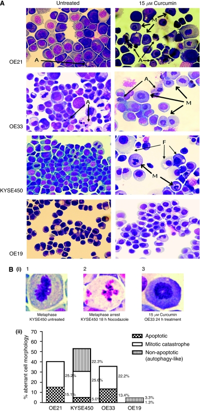
Figure 3.
Morphological features of curcumin-induced cell death in oesophageal cancer cell lines. Cell morphology was visualised using RapiDiff staining by light microscopy after treatment with 15 μM of curcumin for 24 h. (A) Typical cytospin images for untreated and curcumin-treated oesophageal cancer cell lines. Curcumin treatment (15 μM) produced a distinct morphology suggestive of a monopolar spindle and duplicated but unseparated chromosomes centrally located in the cell (M); features are consistent with chromatin images described for mitotic catastrophe (MC). In addition to MC, OE21 and OE33 cell lines show a minor population of cells with clear apoptotic morphology (A). Both of these cell lines also have a background of apoptotic cells visible in their untreated cytospins. By contrast, KYSE450 and OE19 cell lines rarely show apoptotic cells. KYSE450-treated cells predominantly show the typical nuclear morphological features of MC after curcumin treatment. Other features coexist at a background level, including nuclear pyknosis, vacuolisation of the cytoplasm or complete loss of the cytoplasmic membrane while the nuclear membrane remains intact; elements consistent morphologically with autophagic cell death (F). OE19-treated cells preserve their overall structural features at this concentration. (B) (i) Typical chromatin organisations in untreated and treated cells. (1) Normal mitosis metaphase image with chromosomes along the equatorial plane of the cell. (2) Nuclear features of cells treated with 18 h of nocodazole, with typical micronucleation. (3) Chromatin alignment in cells treated with curcumin for 24 h. (ii) Distribution of abnormal morphological features in RapiDiff-stained slides in all four oesophageal cancer cell lines after treatment with 15 μM of curcumin over 24 h. Cells were counted on the basis of their morphology (apoptotic, non-apoptotic/MC and autophagy-like) and expressed as the percentage of the total cell population.

