Figure 5.
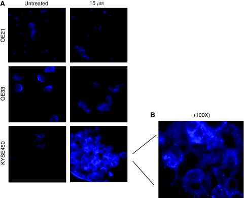
Examination of autophagic cell death in OE21, OE33 and KYSE450 cell lines after treatment with increasing concentrations of curcumin for 24 h. Autophagic vacuoles were identified after incubation of the cells with the selective fluorescent probe monodansylcadaverine (MDC) and visualised immediately by fluorescent microscopy. (A) Fluorescent photographic images of curcumin-sensitive cell lines, OE21, OE33 and KYSE450. Images correspond to untreated and 15 μM of curcumin at 24 h after incubation with MDC. Photographs are representative of two different experiments. Original magnification, × 40. (B) Detailed photograph of the distinct punctate staining in KYSE450 cells after treatment with 15 μM of curcumin and incubation with MDC. Original magnification, × 100.

