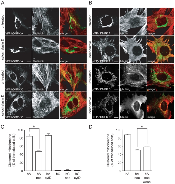
Figure 2. An intact microtubular cytoskeleton enhances hDMPK A–induced perinuclear mitochondrial clustering.
DMPK KO myoblasts were transduced with YFP-hDMPK A or C-expressing adenoviruses in the presence of cytochalasin D (A) or nocodazole (B). F-actin was visualized by fluorescent phalloidin. The microtubular cytoskeleton was stained with an anti-tubulin antibody. Disruption of the actin cytoskeleton did not affect localization of mitochondria. Depolymerization of microtubules decreased mitochondrial clustering in YFP-hDMPK A-expressing cells, but mitochondria still appeared fragmented. The distribution of mitochondria in YFP-hDMPK C-transduced cells was unaffected by nocodazole treatment. Bars, 10 µm. (C) Quantification of mitochondrial clustering. The number of transduced cells that contain clustered mitochondria are expressed as percentage of the total amount of cells expressing hDMPK A at the MOM, with or without treatment of cytochalasin D or nocodazole (images shown in A and B; n = 3, ∼100 cells per experiment, P = 0.01). (D) Effect of nocodazole wash-out. Quantification of the percentage transduced cells with clustered mitochondria after a 12–16 hours treatment with nocodazole, followed by a 8 hours wash-out (n = 3, ∼30 cells per experiment, P<0.05).

