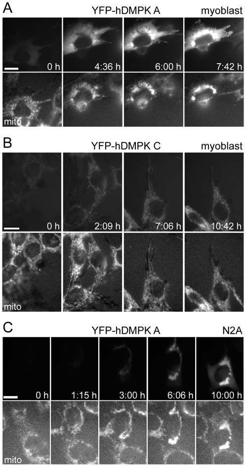
Figure 3. Mitochondrial clustering is a rapid process and occurs already at low hDMPK A expression.
KO myoblasts were transduced with YFP-hDMPK fusion proteins (top panels), while mitochondria stained with MitoTracker Red (bottom panels). Images were collected every three minutes. The time of first appearance of YFP signal was set at t = 0; other time points are indicated in the top panels. (A) YFP-hDMPK A was first detected in the cytoplasm. Soon YFP-hDMPK–decorated mitochondria appeared which then started to cluster, eventually resulting in severely aggregated mitochondria surrounding the nucleus. (B) YFP-hDMPK C directly appeared on mitochondria which maintained their elongated, reticular structure. (C) In N2A cells, YFP-hDMPK A expression emerged in a similar fashion as in KO myoblasts, except that mitochondrial clustering occurred almost immediately and cytosolic staining was less pronounced.

