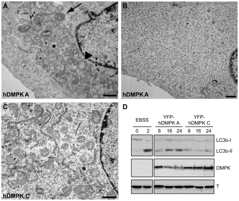
Figure 4. Mitochondrial clustering is accompanied by aberrant ultrastructural morphology.
N2A cells were transfected with YFP-hDMPK A or C isoforms and ultrastructure was analyzed by electron microscopy. (A) YFP-hDMPK A–expressing cells contained fragmented, clustered mitochondria near the nucleus. Cristae structure was often partly lost together with electron density of the matrix (arrowhead) and mitophagy was observed (arrow). In contrast, other areas of the cell were completely devoid of mitochondria (B). (C) Mitochondrial morphology was normal in hDMPK C–expressing cells. A fragment of the nucleus is included here for orientation. Note the proper cristae structure and loose distribution of mitochondria and endoplasmic reticulum. Bars, 1 µm. (D) Lysates from myoblasts expressing YFP-hDMPK A or C for 8, 16 or 24 hours were used for western blotting with a LC3b antibody and showed an increased LC3b conversion following YFP-hDMPK A expression. Cells cultured under normal conditions and nutrient-starved cells, cultured in Earle's Balanced Salt Solution (EBSS) for 2 hours, were used as negative and positive controls, respectively. DMPK expression was verified using a DMPK antibody. Tubulin (T) antibody staining was used as loading control.

