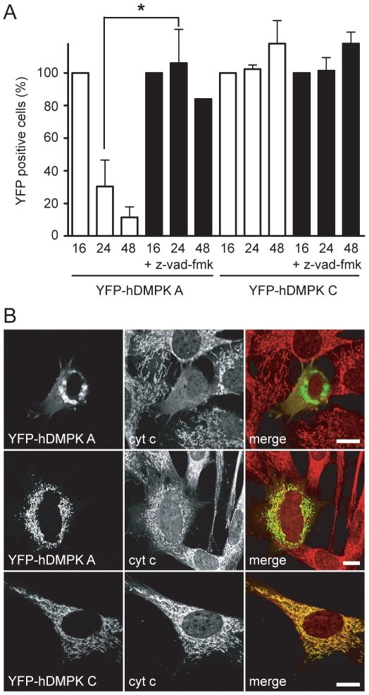
Figure 6. Expression of hDMPK A induces apoptosis.
(A) The effect of apoptosis-inhibitor z-vad-fmk was tested on cell survival of DMPK KO myoblasts transduced with YFP-hDMPK A or C–expressing adenoviruses. YFP-positive cells were counted after 16, 24, and 48 hours. Z-vad-fmk greatly reduced cell death of YFP-hDMPK A–expressing cells, but had no effect when YFP-hDMPK C was expressed. Z-vad-fmk was applied immediately after transduction and maintained present for the remaining time of the experiment. Values at 16 hours were set at 100% (P<0.05, n = 3, >90 cells counted per experiment). (B) YFP-hDMPK A and C–expressing cells were stained for cytochrome c (cyt c). Cells expressing YFP-hDMPK A displayed a diffuse, cytosolic cytochrome c staining when mitochondria were clustered (upper panels). A clear mitochondrial staining was present when mitochondria appeared fragmented (middle panels). A discrete, mitochondrial cytochrome c staining was also observed in YFP-hDMPK C–expressing cells. Bars, 10 µm.

