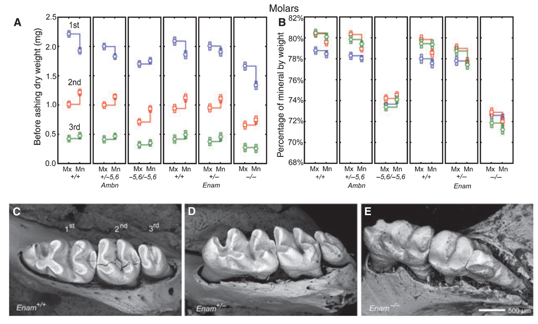
Fig. 4.
Mineral content in intact whole erupted molars. Mean plots ± 95% confidence intervals showing initial dry weight (A) and percentage mineral by weight (B) for the first, second, and third molars removed from wild-type (A, B; Ambn+/+, Enam+/+), heterozygous (A, B; Ambn+/−5,6, Enam+/−), and homozygous (A, B; Ambn−5,6/−5,6, Enam−/−) mice. Panels C–E: scanning electron microscopy images of the first, second, and third mandibular molar crowns from wild-type (C, Enam+/+), heterozygous (D, Enam+/−), and homozygous (E, Enam−/−) mice; cracks within some molar crowns (C, D) are artifacts from freeze drying. The enamel covering the crowns of molars contributes partly to the overall weight of these teeth but is subject to change as molar crowns wear down by occlusal attrition (C,D). Despite this, weight measurements indicate that the enamel covering the molars are affected similarly as incisors, although a gene dosage effect in the molars of heterozygous mice (A, B; Ambn+/−5,6, Enam+/−) is less pronounced than seen on incisors (Fig. 3A,B,E,H). The molars in Enam+/− mice appear to be normal except for some enhanced attrition evident at the distal sides of the second and third molars (D, asterisk). The molars in Enam−/− mice (E) are severely worn down and there is evidence of bone loss around their roots (see Hu et al. (16) for details). Mn, mandibular; Mx, maxillary.

