
Figure 3.
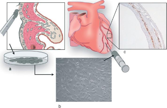
Illustration of EPDC transplantation experiments, (a) Adult epicardial cells are cultured, (b) Cultured epicardial cells are injected into the ischemic area and border zone of the left ventricular wall, (c) Histological section (10×) of the ischemic left ventricular wall after immunohistochemical staining against enhanced green fluorescent protein (eGFP), showing injected eGFP-transduced EPDCs.