
Fig. 3.
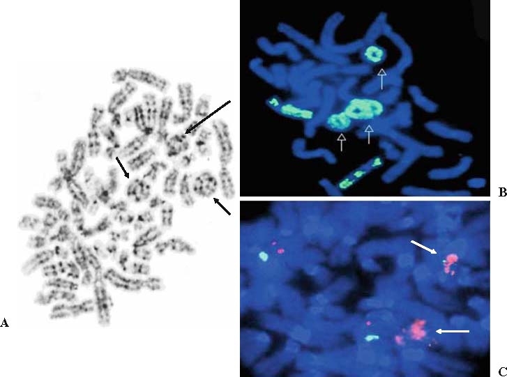
A Multiple ring chromosomes (arrows) in a G-banded metaphase cell of a sclerosing (well-differentiated) liposarcoma. B Ring chromosomes (arrows) are composed of chromosome 12 material, as demonstrated by fluorescence in situ hybridization (FISH) analysis with a whole chromosome 12-paint probe. This image is of a partial metaphase cell also showing two normal chromosome 12 homologues. C Bicolor FISH analysis with a chromosome 12 centromere-specific probe (green) and MDM2 locus-specific probe (red) demonstrates MDM2 amplification in the ring chromosomes (arrows). This image is of a partial metaphase cell also showing two normal chromosome 12 homologues (red and green signals)