
Fig. 6.
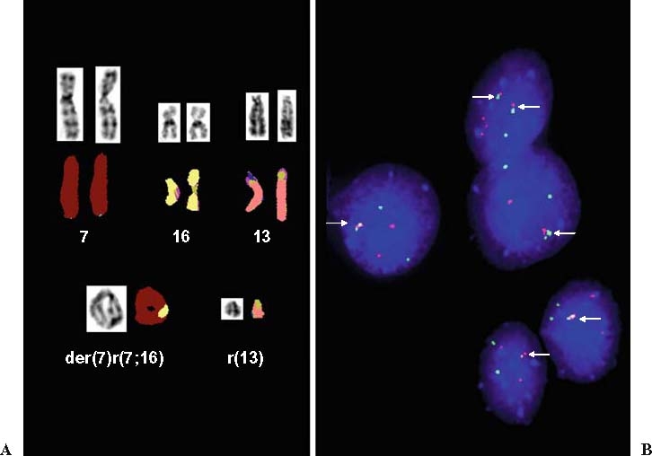
A Partial multifluor (M)-FISH and inverted 4.6-diamino-2-phenylindole (DAPI) image illustrating the chromosomal composition of the two supernumerary ring chromosomes. B Bicolor FISH with a FUS (16p11) spanning probe in Spectrum Green and a CREB3L2 (7q33) spanning probe in Spectrum Red demonstrates fusion of green and red signals (arrows) indicative of the t(7;16)(q33;p11) characteristic of low-grade fibromyxoid sarcoma