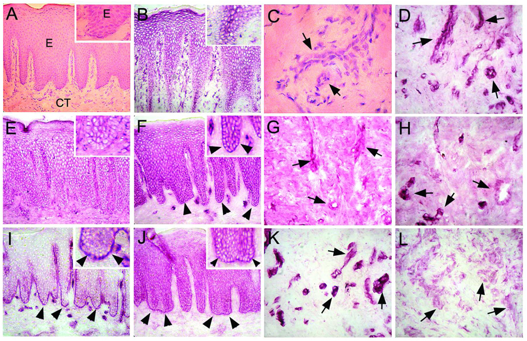
Fig. 1.
Immunolocalization (VIP Staining) of ILK-1 (B, D), kindlin-2 (E, G), migfilin (F, H), paxillin (I, K), and kindlin-1 (J, L) in human palatal oral epithelium and connective tissue. Panels A and C represent hematoxylin-eosin stained sections from the corresponding area. A magnified view of the basement membrane zone (BMZ) and basal cell layer is provided in the top-right corner of the left side panels. ILK-1 (B) and kindlin-2 (E) do not localize to the basement membrane zone or the basal cells, but are found in between cells in the suprabasal layers and blood vessels (D and G, respectively; arrows). Migfilin (F), paxillin (I), and kindlin-1 (J) localize strongly to the basement membrane zone (arrowheads) and between cells throughout all layers of the epithelium with only migfilin (H) and paxillin (K) localizing to blood vessels (arrows). Kindlin-1 (L) expression is weak in blood vessels (arrows).

