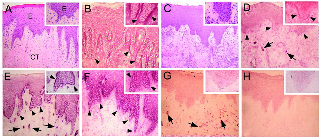
Fig 2.
Immunolocalization (VIP Staining) of migfilin (B, D), paxillin (E, G), and kindlin-1 (F, H) in the oral epithelium of normal (B, E, F) or kindlin-1 deficient gingiva (D, G, H). Panels A and C represents hematoxylin-eosin stained sections from the corresponding areas. A magnified view of the BMZ and basal cell layer is provided in the top-right corner. Migfilin and paxillin fail to localize to the BMZ in kindlin-1 deficient gingiva. Arrowheads point to the BMZ and arrows to blood vessels, respectively.

