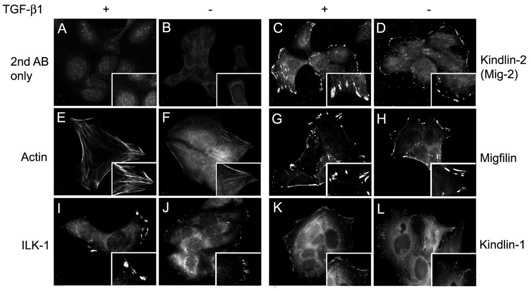
Fig. 3.
Immunolocalization of filamentous actin (E, F) and focal adhesion proteins ILK-1 (I, J), kindlin-2 (C, D), migfilin (G, H), and kindlin-1 (K, L) in HaCaT keratinocytes spread for 16 hours in the presence (A, C, E, G, I, K) or absence (B, D, F, H, J, L) of TGF-β1. Secondary antibody alone (A, B) was used as a negative control. A magnified view of the focal adhesions is provided in the bottom-right corner of each panel. TGF-β promotes expression of actin filaments and an increase in the size of ILK-1, kindlin-2, migfilin, and kindlin-1 focal adhesions.

