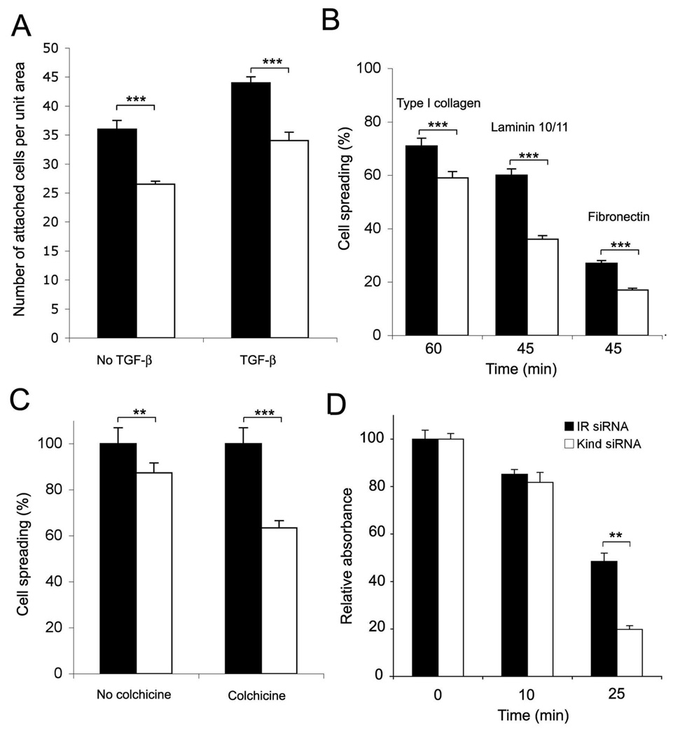
Fig. 6.
Effect of kindlin-1 knockdown on keratinocyte cell adhesion. A, attachment of siRNA treated HaCaT keratinocytes on glass coverslips over 24 hours in the presence or absence of 10 ng/ml TGFβ1; B, spreading of siRNA treated keratinocytes on extracellular matrix proteins (type I collagen, laminin10/11 and fibronectin) for 45–60 minutes; C, spreading of siRNA treated keratinocytes on fibronectinin the presence of absence of 1 µM colchicine. Relative spreading percentage to no drug treatment (control, 100%) is shown; D, detachment of established keratinocyte cultures (confluent siRNA treated cells cultured for 4 days) with diluted (1:4) trypsin at various time points (0–25 minutes). Representative result (mean +/− SEM of triplicate samples) from three parallel experiments is shown (**p<0.01; *** p<0.001).

