
Fig. 1.
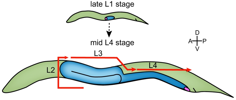
Linker cell migration. By the late L1 stage, the linker cell (LC; pink) has been specified and is positioned to become the leader cell in the male gonad (blue). The LC migrates from the early L2 through the mid-L4 stage. The red arrows indicate the LC migratory path during each of these larval stages. It begins by migrating anteriorly on the ventral bodywall, then turning from the ventral to dorsal side during the L2 molt. It migrates posteriorly during the L3 and L4 stages. In the mid-L3 stage, it performs a second turn from the dorsal back down to the ventral bodywall. In all figures, anterior (A) is left; posterior (P) is right; dorsal (D) is top; ventral (V) is bottom.