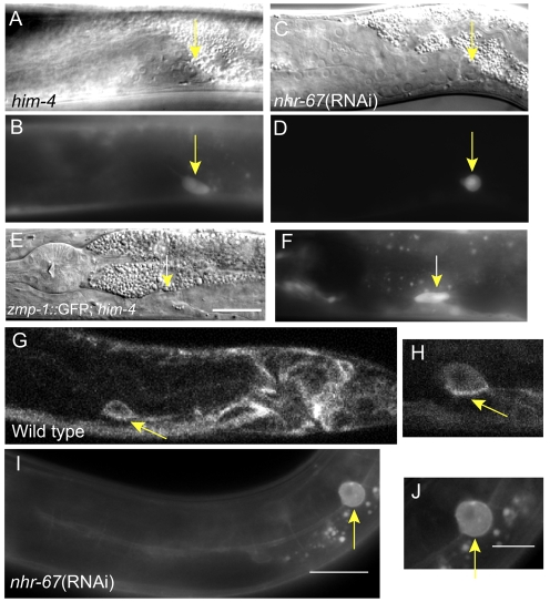
Fig. 5.
Temporal cues are primarily responsible for changes in the LC at the L4 stage. (A-D) The LC in a mid-L4 stage him-4 mutant (A,B) has a polarized shape similar to that of wild-type males (not shown), but different from a similarly positioned LC in an nhr-67(RNAi) male (C,D) of the same stage. (E,F) An L4 stage him-4 mutant correctly expresses zmp-1::GFP in the LC, even though the LC is abnormally positioned near the pharynx (Nomarski, left; fluorescence, right). The LC also has the appropriate elongated morphology of the L4 stage. (G,H) Confocal image of L4 stage male posterior body and a high-magnification image of LC show MIG-2::GFP to be membrane localized to the ventral, adherent side (arrow). (I,J) Nomarski image of nhr-67(RNAi) male showing cytoplasmic and membrane localization of MIG-2::GFP (arrow) in the LC without polarization to the adherent side. Scale bars: 20 μm in E,I; 10 μm in J.

