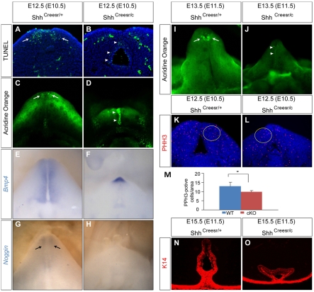
Fig. 2.
Cell death and proliferation analyses in Shh-cKOs. (A,B) TUNEL analysis on coronal sections of GTs (Tm treatment at E10.5) showing distal mesenchymal apoptosis in control (A, arrows) and excessive urethral epithelium (UE) staining in Shh-cKO (B, arrowheads) GT. (C,D) Whole-mount Acridine Orange (AO) staining showing patterned mesenchymal cell death in control (C, arrows), and a drastically augmented UE staining in Shh-cKOs (D, arrowheads). (E-H) Whole-mount in situ hybridization on E12.5 GTs from Shh-cKOs (F,H) and littermate controls (E,G) using the probes indicated. Bmp4 expression is normally detected in the genital mesenchyme (E), and is increased in the distal GT of the mutant (F). Nog is expressed in the distal mesenchyme (arrows in G), and is undetectable in the mutant (H). (I,J) AO staining of E13.5 control (I) and Shh-cKO (J) GT (both were treated with Tm at E11.5) showing a normal mesenchymal staining pattern in the control (I, arrows), and a UE-restricted staining in the Shh-cKO (J, arrowheads). (K-M) Phospho-histone H3 (PHH3) staining on coronal sections of Shh-cKO (L) and controls (K) showing a 20% reduction (M) in PHH3-positive cells in the yellow-circled region. n=8, *P=0.0027. (N,O) Indirect immunofluorescence using a K14 antibody on E15.5 control (N) and Shh-cKO (O) male GT (TM treated at E11.5).

