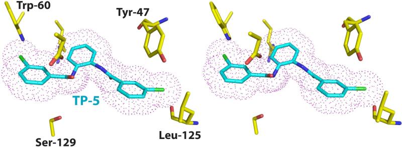
Figure 4. Model of the LasR-TP-5 interaction.
Stereo-view of a model structure of the LasR-TP-5 complex derived from the experimentally determined structure of the LasR-TP-3 complex. The protein residues are shown in yellow and the TP-5 ligand is shown in cyan. A van der Waals surface for the ligand is superimposed and shown as pink dots. Note the steric clash between the backbone carbonyl of Leu-125 and a chlorine atom on the TP-5 ring.

