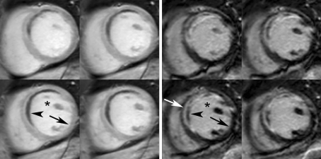
Fig. 3.
Early (left panel) and late CE-CMR images (right panel) in one of the patients. Two sets of four contiguous short axis slices at midlevel of the LV in a patient 5 days after reperfused acute anteroseptal wall myocardial infarction. Left panel 2 min after contrast injection: the blood pool (*) and myocardium show increased signal intensity (arrow), except for a central dark anteroseptal rim (arrowhead; early MVO). Right panel late hyper- and hypoenhancement: 10 min after contrast injection, signal from noninfarcted myocardium is “nulled” (black arrow) and the infarcted myocardium is hyperenhanced (white arrow). A dark central rim is observed within the bright area (late MVO; arrowhead)

