
Figure 1.
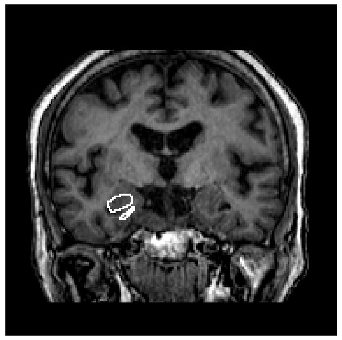
T1-weighted images for acquisition of hippocampus volume. Borders of the hippocampus were traced manually in the coronal orientation with simultaneous monitoring for accuracy in the sagittal and axial orthogonal views.
Official websites use .gov
A
.gov website belongs to an official
government organization in the United States.
Secure .gov websites use HTTPS
A lock (
) or https:// means you've safely
connected to the .gov website. Share sensitive
information only on official, secure websites.
T1-weighted images for acquisition of hippocampus volume. Borders of the hippocampus were traced manually in the coronal orientation with simultaneous monitoring for accuracy in the sagittal and axial orthogonal views.