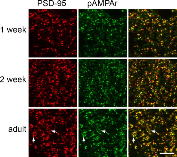
Figure 2.
Colocalization of punctate staining with the PSD-95 and pan-AMPAr (pAMPAr) antibodies in lamina II at different developmental stages. In each row, PSD-95 (red) is on the left, pan-AMPAr (pAMPAr, green) is in the middle, and a merged image is shown on the right. Note that most puncta are stained with both antibodies. Arrows indicate clusters of puncta that probably correspond to synaptic glomeruli. Each image is a projection of two confocal optical sections at 0.3 μm z-spacing. Scale bar, 5 μm.

