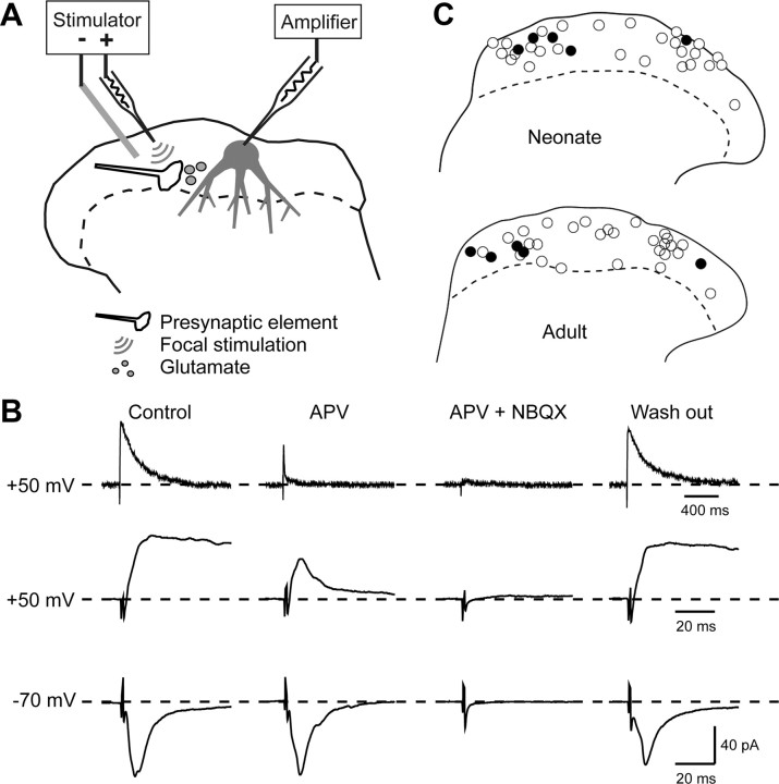
Figure 3.
Recording from superficial dorsal horn neurons and application of focal stimulation. A, Schematic diagram of experimental arrangement. Whole-cell recordings were obtained from lamina I/II neurons, and EPSCs evoked by focal stimulation were recorded while IPSCs were blocked by bicuculline and strychnine. B, Pharmacological demonstrations of EPSC components. At a holding potential of −70 mV (bottom records), inwardly directed EPSCs with fast kinetics were abolished by NBQX but not APV. At a holding potential of +50 mV (top 2 rows of records) outwardly directed EPSCs with slow kinetics were blocked by APV, whereas EPSCs with fast kinetics were sensitive to NBQX. Note different timescales for top and middle rows of records. C, Locations of the intracellularly labeled neurons. The plots show the locations of 32 neurons recorded in slices from neonatal animals (top) and 32 neurons recorded in slices from adult animals (bottom). In each case, cells that showed EPSCs at positive holding potentials are shown with filled circles, and the other cells are shown with open circles.

