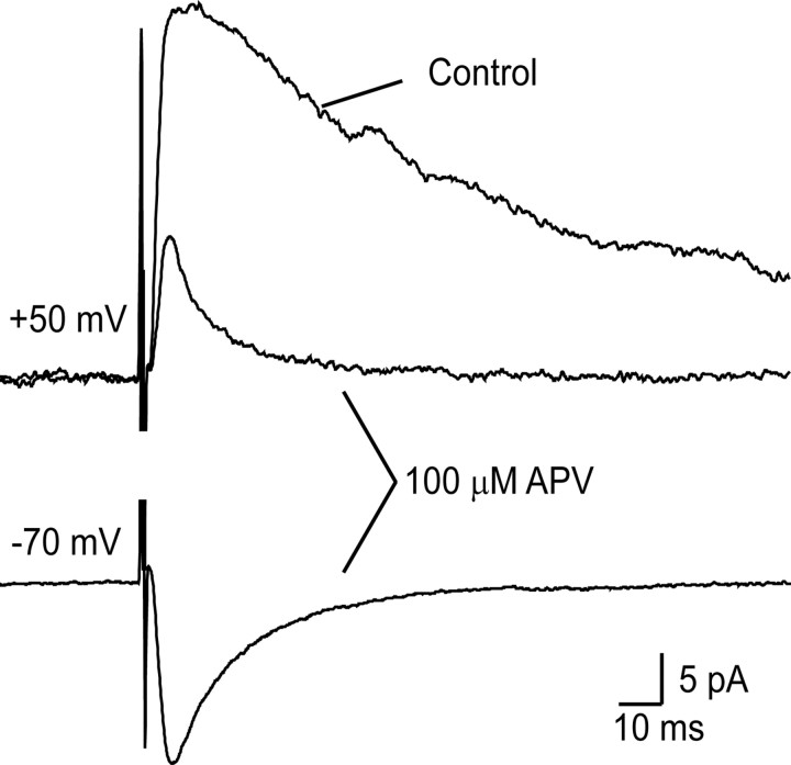
Figure 6.
The effect of APV on EPSCs evoked by focal stimulation while the cell was held at +50 and −70 mV, after reduction of the stimulus strength to below the level that produced EPSCs when the cell was initially tested at −70 mV. The top traces show that APV substantially reduced the EPSC recorded at +50 mV, leaving a small residual current that was presumably mediated by AMPArs. The bottom trace shows a clear EPSC recorded in the presence of APV after the cell was returned to a holding potential of −70 mV. This recording was made in a slice obtained from an adult rat.

