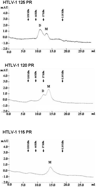
Figure 4. Gelfiltration of the HTLV-1 protease forms.
Gelfiltration of the indicated HTLV-1 protease forms was performed on a Superdex G75 column in 50 mM sodium-acetate buffer, pH 5.0, containing 100 mM NaCl (Buffer B in Table 1), at 0.5 ml/min flow rate. The position of the eluted monomeric (M) and dimeric (D) forms is indicated. Elution volume for the standard proteins used for the calibration of the column under identical conditions is indicated above the chromatograms. Based on the calibration, the apparent molecular weights of the eluted proteins are: HTLV-1 125 PR dimer: 28 kDa, monomer: 12 kDa; HTLV-1 120 PR dimer: 24 kDa, monomer: 11 kDa; HTLV-1 115 PR monomer: 10 kDa.

