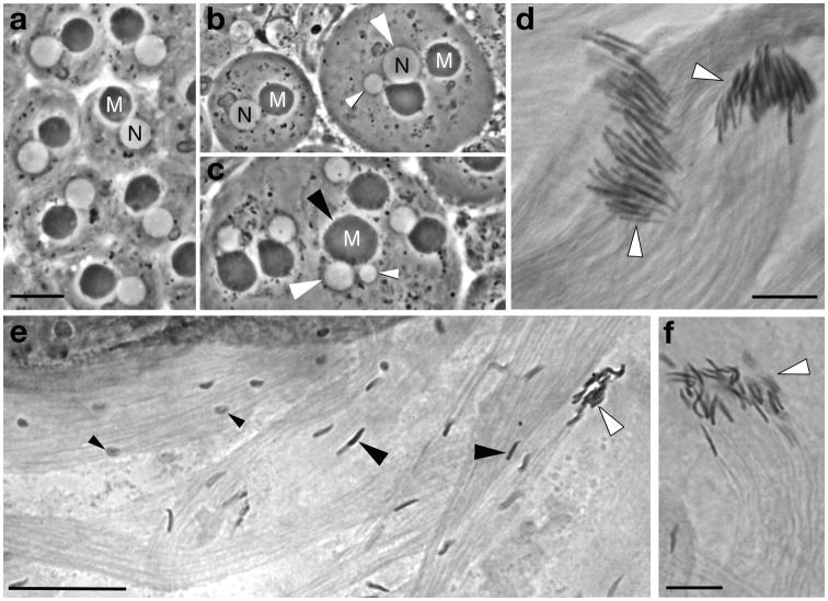
Fig. 3. Meiosis and cytoskeletal microtubule-mediated processes in spermatogenesis in males expressing β1 plus β2 or the paired chimeric β-tubulins with bodies and C-terminal tails swapped.
(a–c) Live young post-meiotic “onion-stage” spermatids with round white nuclei (N) and round (pre-elongation) dark mitochondrial derivatives (M). (a) Spermatids from a fertile male expressing equal amounts of β1 and β2. Spermatids are normal with uniform one to one arrangement of haploid nuclei and normal mitochondrial derivatives. Morphology of spermatids directly reflects meiosis: Spermatid nuclear size indicates nuclear ploidy [Hardy, 1975], and the one-to-one arrangement of nuclei and mitochondrial derivatives of nearly equal size indicates successful cytokinesis [Tokuyasu, 1975]. (b, c) Spermatids in males expressing equal amounts of β1β2C and β2β1C. Most spermatids in most cysts are normal, but some meiotic defects occur in nearly all males of this genotype. (b) The spermatid on the left is normal (compare nuclear and mitochondrial size and arrangement to a). Spermatids on the right have abnormal large and small nuclei (white arrowheads) indicating aneuploidy resulting from defective karyokinesis in meiosis. (c) Two aneuploid nuclei (white arrowheads) associated with a single abnormally large MD (black arrowhead), indicating defective karyokinesis and also failure of cytokinesis at one of the meiotic divisions. (d–f) Elongating spermatid bundles stained with orcein to display sperm nuclei. (d) Spermatid bundles in a male expressing equal amounts of β1 and β2. Spermatids have normal needle-like shaped nuclei that are well aligned at the tips of developing bundles (white arrowheads). The spermatid bundle on the right is slightly more mature than the one on the left, with heads more tightly packed at the tip of the bundle. (e) Spermatids in a male expressing equal amounts of β1β2C and β2β1C. Many nuclei in developing cysts in males of this genotype are poorly shaped and not aligned (small black arrowheads); in addition, many spermatids with shaped nuclei also fail to be correctly aligned (large black arrowheads). Maturing bundles with aligned spermatids with shaped nuclei (white arrowhead) have many fewer spermatids than normal (compare to d). (f) Spermatid bundle (white arrowhead), showing the reduced number of sperm heads resulting from the disruption of alignment (compare to d). Scale bars: 10 μm for (a–c), 10 μm for (d, f), 25 μm for (e).

