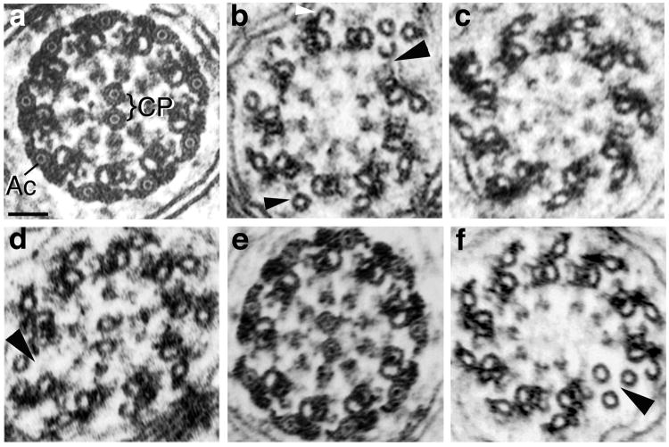
Fig. 4. Axoneme morphology in Drosophila males expressing native and chimeric β-tubulins.
Electron micrographs of axoneme cross-sections. (a) Wild type axoneme morphology in a fertile male expressing equal amounts of β1 and β2. In Drosophila and other insects, in addition to the 9+2 configuration of a ring of nine doublets surrounding a central pair of microtubules (CP), each doublet microtubule has an associated outer accessory microtubule (Ac). In mature axonemes, as shown here, the lumens of the central pair and accessory microtubules contain a luminal filament that appears as a dot in cross-section. (b–d) Typical morphology of intermediate stage axonemes from sterile males expressing transgenic β-tubulins in the absence of β2. (b) Axoneme from a male expressing β1. Central pair microtubules are absent, as is always the case in β1-expressing males. In addition, the ring of doublets is broken (large arrowhead) and some accessory microtubules are not associated with electron dense material (small arrowhead). Incomplete hook-shaped accessory microtubules (white arrowhead) are in the process of assembly [see Raff et al., 1997]. (c) Axoneme lacking central pair microtubules from a sterile male expressing β2β1C. About half of axonemes have central pairs in this genotype (Table II). (d) Axoneme from a sterile male expressing β1β2C. The central pair is present, but the axoneme’s doublet ring is disrupted (large arrowhead). About half of axonemes have central pairs in this genotype (Table II). (e, f) Examples of the range of axoneme morphology in sterile or weakly fertile males expressing one copy β1β2C and one copy β2β1C, in the absence of β2. (e) Nearly mature axoneme with wild type morphology (compare with a). (f) Intermediate stage axoneme lacking central pair microtubules. The doublet ring is also broken (arrowhead). Scale bar in (a) for all panels: 50 nm.

