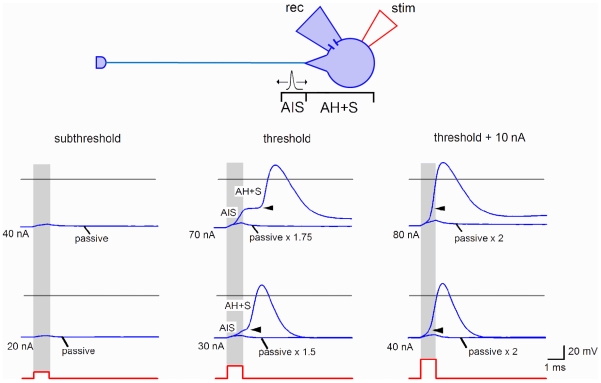
Figure 3. Spike initiation in an SG neuron.
Stimulating (cell-attached) and recording (perforated-patch) pipettes were placed on the soma of an SG neuron. Stimulation (1 ms) increased with a 10 nA increment. Subthreshold, the membrane responses were considered as passive. Threshold, the AIS and AH+S components could be best distinguished (transitions are indicated by arrowheads). The AIS component was initiated within the 1 ms of the stimulation. The passive response was multiplied by the corresponding scaling factor. Threshold+10 nA, the interval between the AIS and AH+S components became shorter. The AIS component was completely generated within the 1 ms of the stimulation. Two SG neurons shown have different proportions between the amplitudes of the AIS and AH+S components and different kinetics of transitions. Horizontal grey lines, 0 mV. Resting potentials were about −70 mV.

