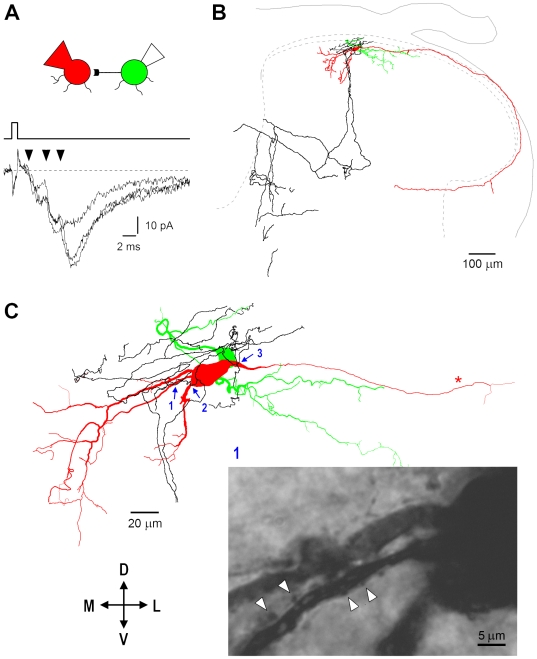
Figure 15. Reconstruction of synaptically connected neurons.
A, composite EPSCs (−80 mV) with at least three components (arrowheads). The postsynaptic neuron was filled with biocytin in whole-cell mode, while the presynaptic one through the cell-attached pipette [28]. B, both cell bodies were in the SG. The axon of the postsynaptic neuron (red) ran along the dorsal surface of the grey matter, gave a collateral in the lateral column, and turned medially to re-enter the grey matter. The presynaptic cell axon (black) formed a dense network in the vicinity of the cell bodies and divided into two major branches which travelled to deeper laminae and turned towards the dorsal grey commissure. Soma and dendrites of the presynaptic neuron are shown in green. C, a number of close appositions between the varicosities of the presynaptic axon and the dendrites and soma of the postsynaptic neuron were detected in regions 1–3 (blue arrows). Some of them are shown (arrowheads) on the photomicrograph of the region 1. Objective; 100x (oil-immersion), numerical aperture 1.25.

