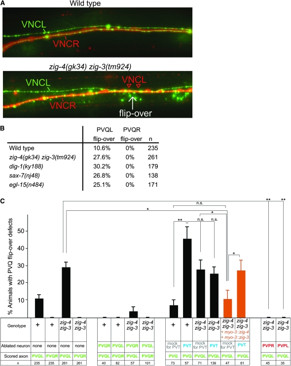
Figure 6.—
Cellular analysis of axon midline flip-over defects. (A) Midline flip-overs are all-or-none defects as assessed by colabeling the entire left and right ventral nerve cord fascicle with a panneuronal rfp marker (otIs173) and the PVQ neurons with a gfp marker (oyIs14). Young adult animals are shown. (B) PVQ axon flip-overs (scored in young adults with the oyIs14 and otIs173 transgenes) always occur from the left into the right fascicle, as scored in different maintenance factor mutant backgrounds. This also holds for the background level of flip-over in a wild-type genetic background. (C) Laser ablation data. “+” = wild-type strain; “zig-4 zig-3” = zig-3(tm924) zig-4(gk34). For the PVT ablation experiments, a gcy-1∷gfp transgene was placed into the genetic background to visualize PVT. PVQ axons and soma were visualized with oyIs14 and PVP axons and soma with hdIs26. Proportions of different animal populations were compared using the z-test. *P < 0.05; **P < 0.01; and NS: P > 0.05.

