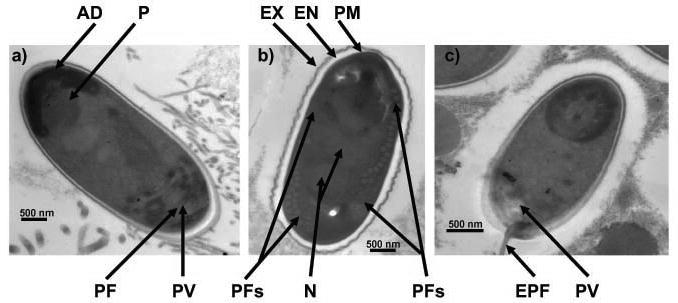
Fig. 6--8.

Electron-micrographs of longitudinal sections of spores of Nosema ceranae. 6. Micrograph showing anchoring disk (AD), polaroplast (P), posterior vacuole (PV), polar filament (PF). 7. Micrograph showing endospore (EN), exospore (EX), plasmamembrane (PM), nucleus (N), 20--22 isofilar coils of the polar filament (PFs). 8. A spore with an extruded polar filament (EPF). Note the more conspicuous PV.
