
Fig. 4. Targeted therapy of spontaneous popliteal lymph node metastasis with S. typhimurium A1-R.
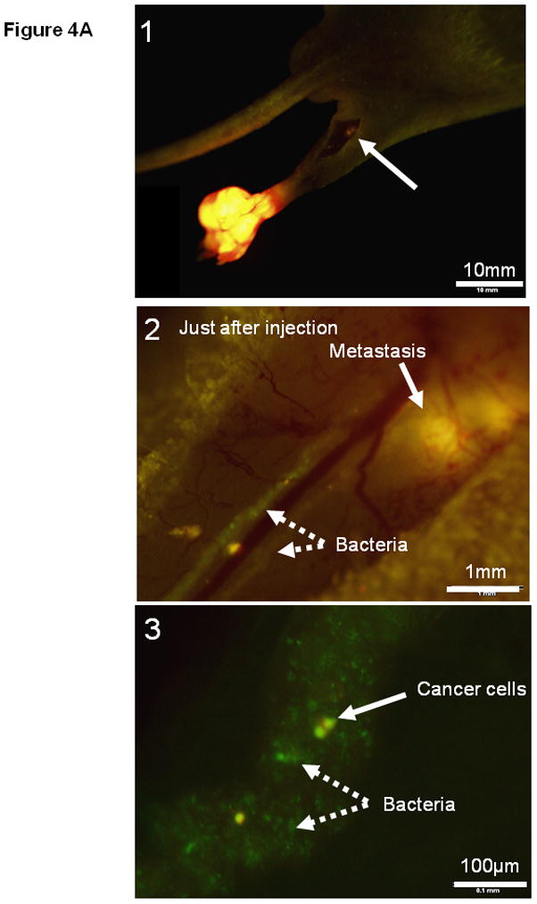
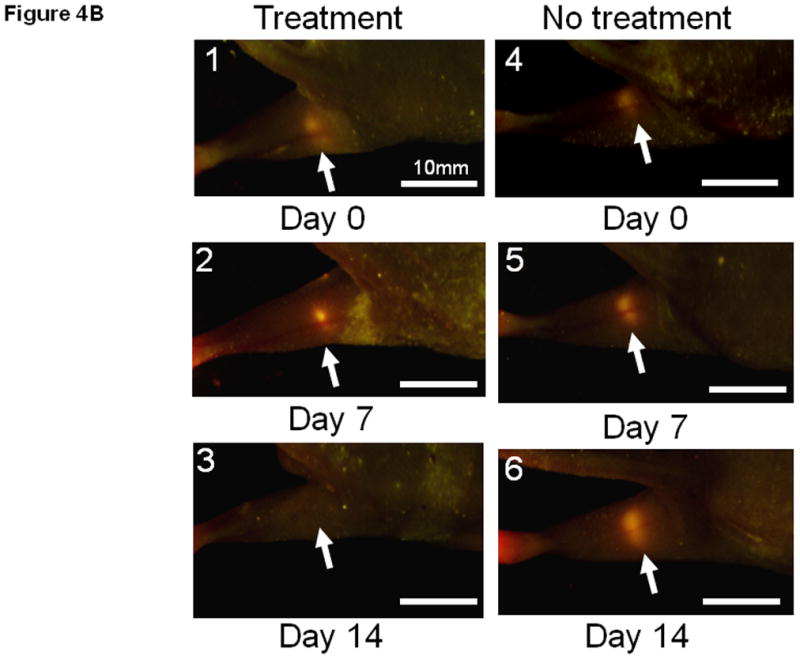
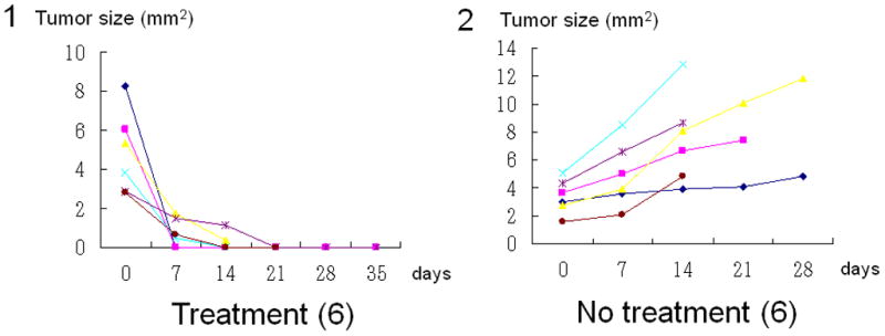
(A1) Dual color HT-1080 human fibrosarcoma cells were injected into the foot pad in nude mice. The popliteal lymph node metastasis was imaged every week after cancer-cell injection. Once metastasis was visualized in the popliteal region (arrow), bacterial therapy was started to target the metastasis. (A2) Bacteria were injected subcutaneously in the foot pad. The popliteal lymph node was exposed to image bacteria trafficking. GFP bacteria were observed in lymphatic channels connecting the popliteal lymph node. (A3) Higher magnification of lymphatic channel. GFP bacteria and dual color cancer cells are readily distinguished. (B1–B6) Representative weekly images of the popliteal lymph node metastasis after bacteria injection. (B1–B3) 0, 7 and 14 days after bacteria injection. The lymph node metastasis has been eradicated by 14 days. (B4–B6) 0, 7 and 14 days for control (no bacteria treatment). The lymph node metastasis continues to grow. (C1–C2) All lymph node metastases have regressed and 5 out of 6 are eradicated within 7 to 21 days after treatment in contrast to growing tumors in the control group.