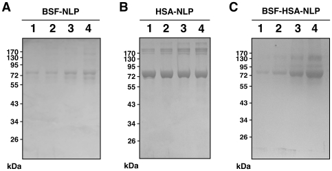
Figure 8. Differences in the binding affinities between fetuin-A and albumin toward NB-like particles as revealed by SDS-PAGE.
NB-like particles were prepared from 3 mM each of CaCl2 and NaH2PO4 in 1 ml of DMEM containing either BSF, HSA, or both proteins at the following concentrations: 20 µg/ml, 40 µg/ml, 80 µg/ml, or 160 µg/ml of BSF, corresponding respectively to lanes 1–4 in (A); 0.2 mg/ml, 0.4 mg/ml, 0.8 mg/ml, and 1.6 mg/ml of HSA, respectively, lanes 1–4 in (B); or both proteins at these same concentrations for lanes 1–4 in (C), respectively. Following incubation in cell culture conditions for 1 month, the NB-like particles were pelleted by centrifugation, washed twice in HEPES buffer, resuspended in 50 µl of 50 mM EDTA, and processed for SDS-PAGE as described in the Materials and Methods . In the case of BSF-NLP shown in (A), fetuin-A appeared as a major band slightly above 72 kDa (lane 1), with additional bands of higher and lower molecular weights noticeable at higher protein concentrations (lanes 2 to 4). In HSA-NLP (B), albumin formed a major band of strong intensity at 72 kDa. Note a higher molecular band above the 170 kDa marker that appears to increase steadily from lanes 1 through 4, while the 72 kDa band in the 4 lanes appears to decrease slightly in intensity from left to right. In the case of BSF-HSA-NLP (C), note the increase in the intensity of bands at 72 kDa and at a higher molecular weight. The bands of higher molecular weights may be due to altered migration or aggregation of the purified proteins used.

