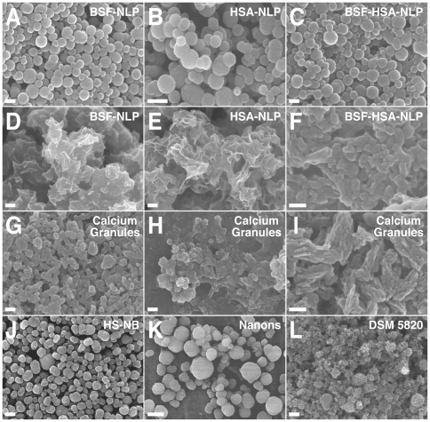
Figure 9. Protein-mineral nanoparticles seeded by fetuin-A and albumin show morphological resemblance to NB and calcium granules when observed by SEM.
Protein-mineral nanoparticles were prepared by adding 0.3 mM of CaCl2 and NaH2PO4 to DMEM containing BSF at 2.1 µg/ml (A and D, labeled as “BSF-NLP”), HSA at 120 µg/ml (B and E, “HSA-NLP”), or both BSF and HSA at these same concentrations (C and F, “BSF-HSA-NLP”), followed by incubation for either 3 days (A–C) or 1 month (D–F) in cell culture conditions and preparation of the particles for TEM, as described in the Materials and Methods . Protein-mineral nanoparticles containing BSF and/or HSA showed a round morphology when observed after 3 days (A–C), but they tended to coalesce to form films or aggregates when incubated for a longer period of 1 month (D–F). Note the presence of structures resembling cells undergoing cell division in (B). Calcium granules were prepared from the addition of either CaCl2 (G), NaH2PO4 (H), or a combination of both (I) to FBS, followed by overnight incubation, centrifugation, and the washing steps described in the Materials and Methods . Calcium granules showed variable morphologies, consisting of either round particles (G) or film/aggregate-like structures (H and I). The morphologies of both the protein-mineral nanoparticles and the calcium granules were similar to NB obtained from 10% HS (J, “HS-NB”) or the NB strains “Nanons” (K) and “DSM 5820” (L) which were both maintained in 10% FBS. These NB samples revealed either round particles (J and K) or more crystalline structures harboring elongated crystal projections and aggregates (L). Scale bars: 100 nm (F, I); 200 nm (A, C–E, G, H, J, L); 400 nm (B); 1 µm (K).

