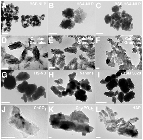
Figure 10. Protein-mineral nanoparticles containing fetuin-A and albumin resemble NB and calcium granules as revealed by TEM.
Protein-mineral nanoparticles were prepared as described in Fig. 9, from solutions containing either BSF (A), HSA (B), or both proteins (C), followed by the addition of 0.3 mM each of CaCl2 and NaH2PO4, and incubation for 1 month in cell culture conditions. The samples were then prepared for TEM without fixation or staining. The various protein-seeded particles showed small, rounded formations that tended to aggregate (A–C). The samples of calcium granules prepared from addition of either CaCl2 (D), NaH2PO4 (E), or a combination of both (F) to FBS (see Materials and Methods ) showed stacks of crystalline spindles and stacks forming ellipsoid structures (D and E), while other granules appeared mostly as round aggregated particles (F). The morphologies of the protein-mineral nanoparticles and the calcium granules were similar to NB obtained from either 10% HS (G, “HS-NB”) or the two NB strains “Nanons” (H) and “DSM 5820” (I) which were both obtained from 10% FBS. In this case, the NB samples consisted of rounded particles with rough surfaces and variable sizes (G–I). Commercially available CaCO3 (J), Ca3(PO4)2 (K), and HAP (L) incubated in DMEM for 1 hour showed mainly large, crystalline, monolithic structures. Scale bars: 50 nm (A, F); 100 nm (B, D, E, G–J, L); 200 nm (C, K).

