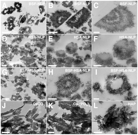
Figure 11. Thin-sections of protein-mineral nanoparticles seeded by fetuin-A or albumin show distinct morphologies.
Protein-mineral nanoparticles were prepared as described in Fig. 6, by diluting either BSF at 2.1 µg/ml (A–C), HSA at 120 µg/ml (D–F), or both proteins at these same concentrations (G–I) into DMEM and then adding 0.3 mM each of CaCl2 and NaH2PO4, followed by incubation in cell culture conditions for 1 month. Thin-sections were prepared without fixation or staining, as described in the Materials and Methods . BSF-NLP (A–C) resembled multi-layered laminations with alternate electron densities. HSA-NLP (D–F) appeared mostly as incompletely sealed, single-layered formations. The BSF-HSA-NLP particles had rough surfaces covered with elongated crystal projections (G), with some structures appearing either as multi-layered (H) or single-layered (I). Control, commercial grades of CaCO3 (J), Ca3(PO4)2 (K), and HAP (L) were incubated in DMEM as in Fig. 10. These controls showed mainly monolithic platelets or aggregates of crystalline formations that contrasted with the round nanoparticles shown above. Scale bars: 100 nm (H, I, L); 200 nm (C, K); 250 nm (F); 500 nm (B, D, E, G, J); 1 µm (A).

