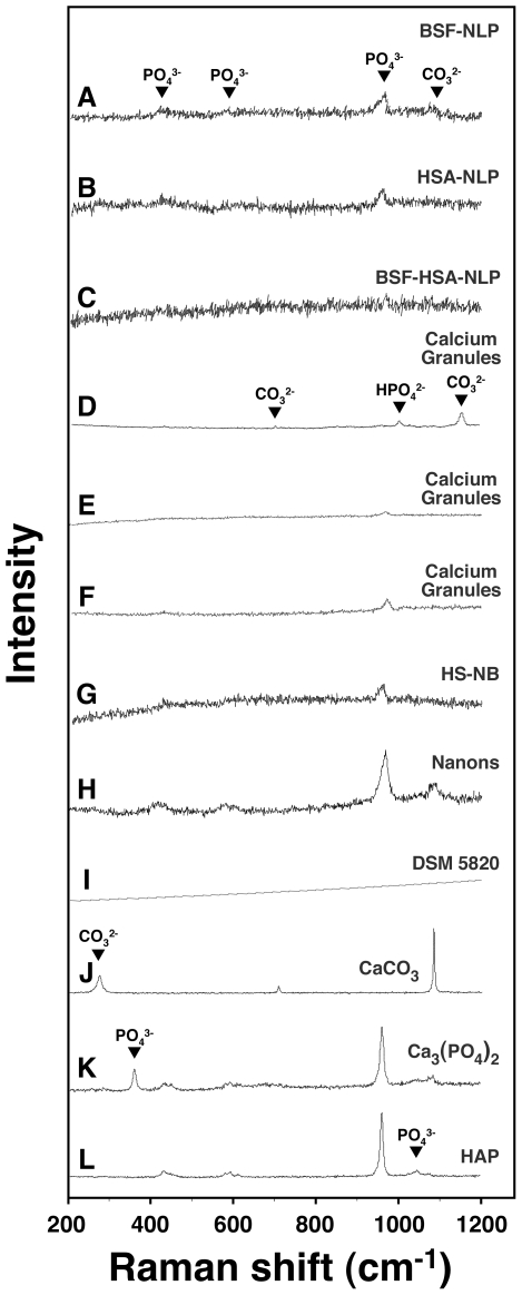
Figure 14. Micro-Raman spectroscopy of protein-mineral nanoparticles shows chemical compositions similar to those of calcium granules and NB.
Protein-mineral nanoparticles were prepared as in Fig. 9, by adding 0.3 mM each of CaCl2 and NaH2PO4 to DMEM containing BSF (A), HSA (B), or both proteins (C), followed by incubation in cell culture conditions for 1 month and processing for micro-Raman spectroscopy. Calcium granules were obtained by adding CaCl2 (D), NaH2PO4 (E), or both (F) to FBS, followed by overnight incubation and preparation for micro-Raman spectroscopy as described in the Materials and Methods . Micro-Raman spectra were also acquired for NB that were initially cultured from 10% HS (G, “HS-NB”) or 10% FBS (H and I, “Nanons” and “DSM 5820”, respectively). These nanoparticle samples showed phosphate groups at 361 cm−1, 440 cm−1, 581 cm−1, 962 cm−1, 1,002 cm−1 (HPO4 2−), and 1,048 cm−1 and carbonate moieties at 280 cm−1, 712 cm−1, 1,080 cm−1, and 1,150 cm−1. The protein-mineral nanoparticles mainly showed peaks of phosphate and lower peaks of carbonate (A–C) while the calcium granules (D–F) and NB (G–I) samples showed carbonate and phosphate peaks of variable intensities. The three controls CaCO3 (J), Ca3(PO4)2 (K), and HAP (L), diluted and washed in double-distilled water, were included for comparison.

