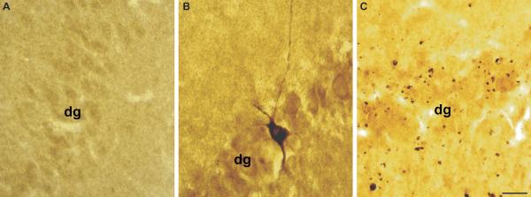
Figure 4.
Calpain-mediated alpha-II-spectrin (CMSP) proteolysis in the hippocampus following diffuse brain injury. Immunostaining for the 145-kDa breakdown product of alpha-II-spectrin demonstrates enhanced levels of spectrin proteolysis in the hippocampus. (A) Little to no SBDP145 immunoreactivity is observed in the hippocampus of sham animals. (B) Isolated neuronal somatic SBDP145-immunoreactivity is seen in the granule cell layer of the dentate gyrus at 3 hours post-injury. (C) Immunostaining is widespread throughout the hippocampus at 24 hours and later time points post-injury (not shown). Staining is primarily localized to axons and axonal debris. dg = dentate gyrus. Scale bar = 50 μm

