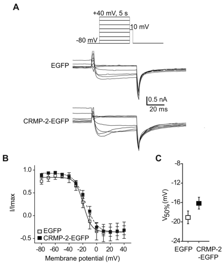
Fig. 5.
Effect of CRMP-2 on DRG Cav2.2 inactivation. (A) Top panel: voltage protocol. Currents were evoked by voltage steps from –80 to +40 mV in 10-mV increments prior to delivering a test pulse to +10 mV. Bottom two panels: exemplar current traces from EGFP and CRMP-2–EGFP transfected neurons. (B) Normalized test pulse peak current amplitude plotted against its preceding holding potential and fitted with the Boltzmann relation. (C) Plot of mean V50% for EGFP and CRMP-2–EGFP showed no difference between the groups.

