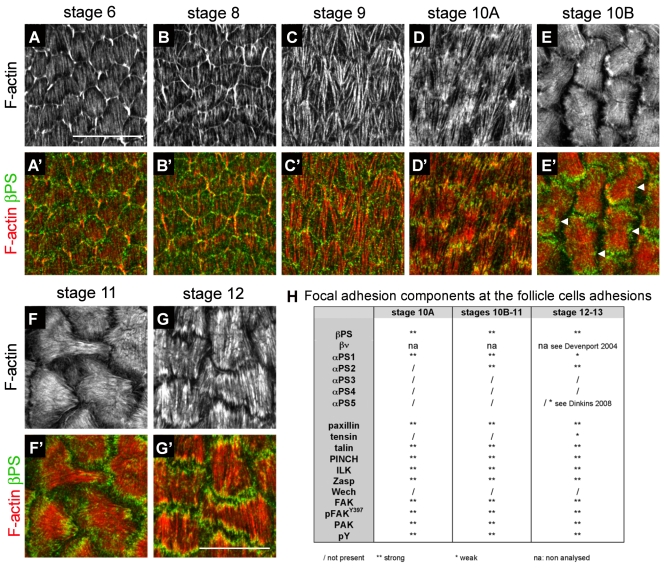
Fig. 2.
Time course of stress-fibre organization throughout oogenesis and summary of the distribution of all focal-adhesion molecules tested. (A-G′) Micrographs of basal actin stress fibres from stage 6 to stage 12, showing F-actin (A-G) and its merge with βPS (A′-G′). All images are oriented posterior to the right. Basal F-actin is present from the beginning of oogenesis (low staining in cell middles in A), but new filaments become visible from stage 6 (high staining originating from cell borders and pointing down). These new filaments extend at stage 8 (B) and, together with the early filaments, form long fibres covering the cells at stage 9 (C). These fibres are more apparent at stage 10A (D). Up to stage 10A, the fibres orient perpendicular to the long axis of the egg. At stage 10B, the fibres thicken (E) and change their orientation by 90° by stage 11 (F), adopting a fan shape. At stage 12, the original orientation is recovered and stress fibres are striated (G). βPS localizes at actin-fibre ends at all stages, and also at cell borders. (E′) Note that a central patch of βPS is also present, suggesting de novo adhesion (arrowheads). (H) Table summarizing all focal-adhesion components tested and their presence at stages 10A, 11 and 12. Scale bars: 20 μm.

