
Figure 2.
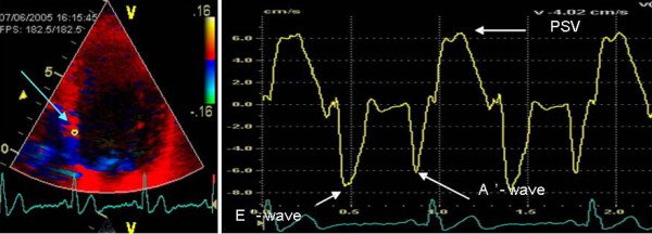
Tissue Doppler echocardiography. To the left, an apical four-chamber view picture with the sample volume placed at the basal portion of the septal wall. On the right, all the variables extracted from on-line recording.
Official websites use .gov
A
.gov website belongs to an official
government organization in the United States.
Secure .gov websites use HTTPS
A lock (
) or https:// means you've safely
connected to the .gov website. Share sensitive
information only on official, secure websites.
Tissue Doppler echocardiography. To the left, an apical four-chamber view picture with the sample volume placed at the basal portion of the septal wall. On the right, all the variables extracted from on-line recording.