Figure 6.
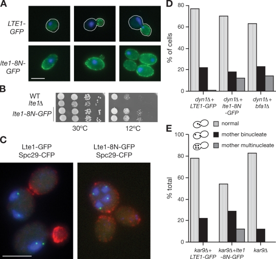
Lte1-8N is localized at mother and daughter cell cortexes and abrogates the SPoC. (A) Subcellular localization of wild-type and 8N forms of Lte1 expressed in SY144 lte1Δ in unbudded cells (left), cells with a small bud (middle), and large-budded cells (right). Cells are outlined in white. (B) Complementation of the cold sensitivity of lte1Δ mutants by lte1-8N–GFP. WT, wild type. (C) SPoC activity in MGY407 (LTE1-GFP dyn1) and MGY408 (lte1-8N–GFP dyn1) cells expressing SPC29-CFP grown overnight at 14°C. The left panel shows examples of LTE1 cells with binucleate mother cells, and the right panel shows lte1-8N mutants with multiple nuclei in the mother and multiple anucleate buds. Lte1 is shown in red, Spc29 is shown in green, and DNA is shown in blue. (D) The frequencies of normal and bi- and multinucleate cells in C (n > 100). (E) SPoC activity at 30°C in logarithmically growing MGY459 (kar9Δ), MGY462 (kar9Δ LTE1-GFP), and MGY463 (kar9Δ lte1-8N–GFP) cells expressing Spc29-CFP. The histogram shows frequencies of normal and bi- and multinucleate cells (n > 100). Bars, 5 µm.

